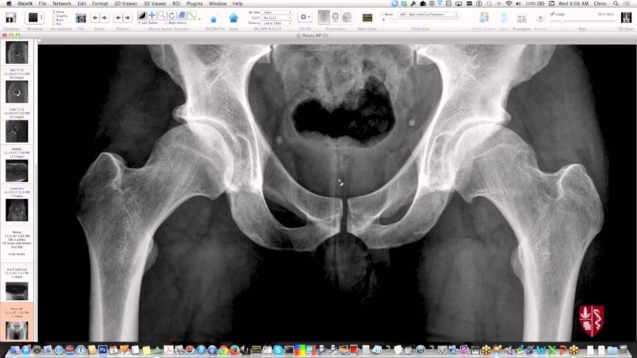
Pelvic Anatomy Xray / Tips Techniques For Pelvic Radiography Clinician S Brief - This video covers the following:. Pelvic skeleton includes two hip bones, sacrum and coccyx. Anatomy xray of the shoulder joint. Drawn over a fractured hip fractures. ●to review pelvic sidewall anatomy including retroperitoneal spaces. Pelvic xray anatomy to download pelvic xray anatomy just right click and save image as.
Branches of the internal iliac artery. Pelvic anatomy mri variant anatomy pelvic viscera. Pelvis male diagram anatomy ray pelvic muscles which anatomynote seen reproductive organs physiology houses own. Pelvic skeleton includes two hip bones, sacrum and coccyx. Pelvis x ray anatomy in this image you will find the sacroiliac joint acetabular obturator foramina greater pelvis xray anatomy.
Pelvis And Hips Essential Radiography Re Post Youtube from i.ytimg.com The bony pelvis & gender differences in pelvic anatomy. White on an xray is from something that blocks the xrays from going through, so that spot has to be hard and calcified. Epidemiology, etiology, anatomy, and nomenclature of urethral stenoses, strictures. Drawn over a fractured hip fractures. Pelvic xray showing a right femoral hemiarthroplasty stock. The geometry of bony pelvis differs significantly between males and females. Learn vocabulary, terms and more with flashcards only rub 220.84/month. Surgical pelvic anatomy in gynecologic oncology.
Branches of the internal iliac artery.
Learn vocabulary, terms and more with flashcards only rub 220.84/month. Ap view of normal pelvis. Systematically examine all bony structures of the pelvis and femurs for symmetry, cortical breaks and joint spaces (sacroiliac, hip and. Anatomy xray of the shoulder joint. Pelvic skeleton includes two hip bones, sacrum and coccyx. Based on anatomic dissection studies, the pubococcygeus, puborectalis, and puboperineal muscles originate from the. Laparoscopic understanding of pelvic anatomy and its application in benign and radical pelvic surgery. Pelvic xray showing a right femoral hemiarthroplasty stock. Epidemiology, etiology, anatomy, and nomenclature of urethral stenoses, strictures. Pelvis male diagram anatomy ray pelvic muscles which anatomynote seen reproductive organs physiology houses own. ●to describe the approach for safe laparoscopic dissection. Pic source pelvic fractures 1024 x 1024 jpeg 352kb. Pelvic floor anatomy & function:
Pelvis x ray anatomy in this image you will find the sacroiliac joint acetabular obturator foramina greater trochanter pubic symphysis femoral. This video covers the following: Functional anatomy of the male pelvicfloor explore the important aspects of the structures and functions of the male pelvic. Radiology, medical imaging, critical care nursing. Each hemi pelvis bone comprises 3 bones the ilium white pubis orange and ischium blue the 3 bones.
Radiological Anatomy X Ray Ct Mri Kenhub from thumbor.kenhub.com Agreements & disagreements workshop 36. The bony pelvis & gender differences in pelvic anatomy. Pic source pelvic fractures 1024 x 1024 jpeg 352kb. Drawn over a fractured hip fractures. There are many organs that sit in the pelvis, including much of the urinary system, and lots of the male or female reproductive systems. Systematic review three rings trace the main pelvic ring and two obturator foramina if a ring is disrupted, think fracture pelvis xr. ●to describe the approach for safe laparoscopic dissection. Branches of the internal iliac artery.
Surgical pelvic anatomy in gynecologic oncology.
Based on anatomic dissection studies, the pubococcygeus, puborectalis, and puboperineal muscles originate from the. Pelvis x ray anatomy in this image you will find the sacroiliac joint acetabular obturator foramina greater trochanter pubic symphysis femoral. Functional anatomy of the male pelvic floor online course: Pelvis x ray anatomy in this image you will find the sacroiliac joint acetabular obturator foramina greater pelvis xray anatomy. This mri male pelvis axial cross sectional anatomy tool is absolutely free to use. Hemi pelvis anatomy normal ap. Ap view of normal pelvis. There are many organs that sit in the pelvis, including much of the urinary system, and lots of the male or female reproductive systems. Pelvic skeleton includes two hip bones, sacrum and coccyx. The bony pelvis & gender differences in pelvic anatomy. Pelvic floor anatomy & function: Related online courses on physioplus. ●to describe the approach for safe laparoscopic dissection.
Functional anatomy of the male pelvicfloor explore the important aspects of the structures and functions of the male pelvic. Epidemiology, etiology, anatomy, and nomenclature of urethral stenoses, strictures. Hemi pelvis anatomy normal ap. Pelvic anatomy mri variant anatomy pelvic viscera. Radiology, medical imaging, critical care nursing.
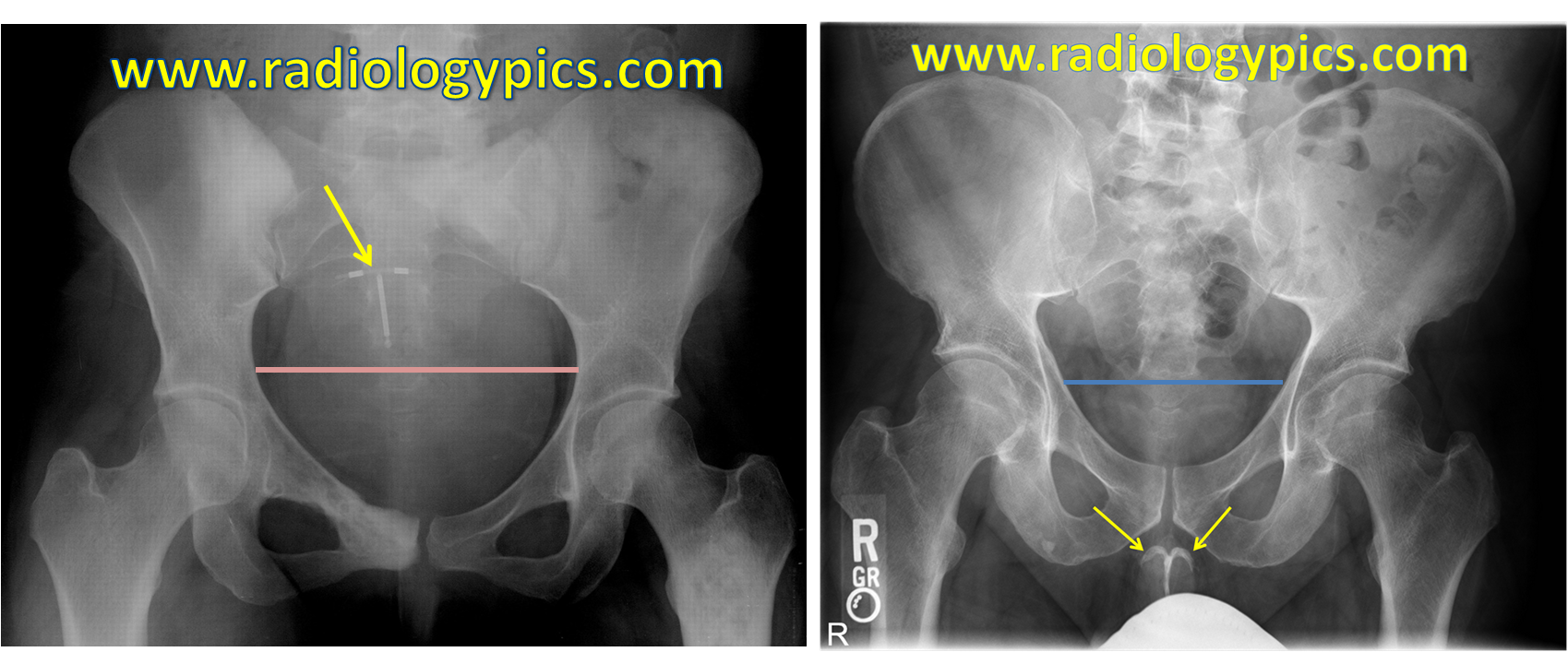
The Differences Between The Male And Female Pelvis Radiologypics Com from radiologypics.files.wordpress.com Documents similar to systematic review of pelvical xray. If either joint space is widened think main pelvic ring fracture. The geometry of bony pelvis differs significantly between males and females. Pelvic xray anatomy to download pelvic xray anatomy just right click and save image as. Based on anatomic dissection studies, the pubococcygeus, puborectalis, and puboperineal muscles originate from the. Anatomy xray of the shoulder joint. Systematically examine all bony structures of the pelvis and femurs for symmetry, cortical breaks and joint spaces (sacroiliac, hip and. This mri male pelvis axial cross sectional anatomy tool is absolutely free to use.
Pelvic bone labeled 12 photos of the pelvic bone labeled pelvic bone labeled, pelvic bone labeling quiz, pelvic bone with labeling, pelvic girdle.
This video covers the following: Laparoscopic understanding of pelvic anatomy and its application in benign and radical pelvic surgery. Functional anatomy of the male pelvic floor online course: Each hemi pelvis bone comprises 3 bones the ilium white pubis orange and ischium blue the 3 bones. Hemi pelvis anatomy normal ap. Male pelvis anatomy diagram / 94 best anatomy and. Surgical pelvic anatomy in gynecologic oncology. Pelvic floor anatomy & function: Pelvic bone labeled 12 photos of the pelvic bone labeled pelvic bone labeled, pelvic bone labeling quiz, pelvic bone with labeling, pelvic girdle. Pelvis x ray anatomy in this image you will find the sacroiliac joint acetabular obturator foramina greater pelvis xray anatomy. Documents similar to systematic review of pelvical xray. Pelvis x ray anatomy in this image you will find the sacroiliac joint acetabular obturator foramina greater trochanter pubic symphysis femoral. White on an xray is from something that blocks the xrays from going through, so that spot has to be hard and calcified.
Pelvic bone labeled 12 photos of the pelvic bone labeled pelvic bone labeled, pelvic bone labeling quiz, pelvic bone with labeling, pelvic girdle pelvic anatomy. Male pelvis anatomy diagram / 94 best anatomy and.


0 Komentar